Heterotopic bone formation, also known as heterotopic ossification (HO), is a medical condition where bone tissue forms outside the skeletal system, often within muscles or soft tissues.
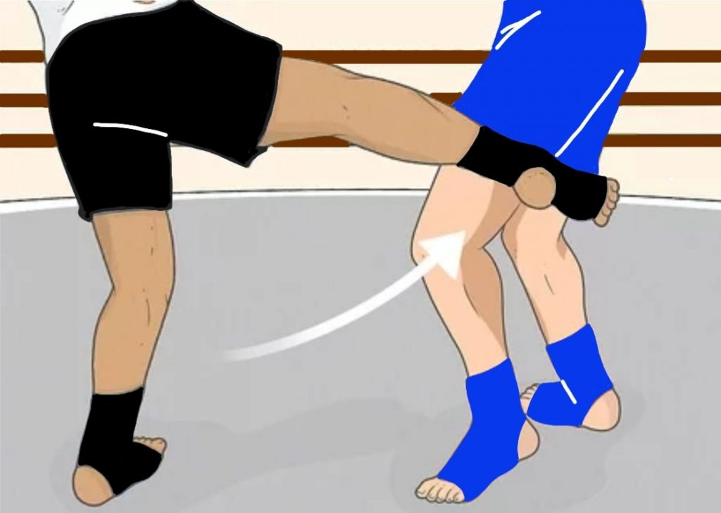
This abnormal bone growth can occur after traumatic injuries, surgeries, or severe burns. One potential cause is heterotopic bone formation resulting from traumatic blows, such as forceful kicks to the thigh, a technique commonly used in Muay Thai and full contact Karate, delivered with the shin.
A hard kick to the thigh can result in significant tissue damage, including muscle contusion and hematoma formation. The body’s natural response to such an injury is inflammation, followed by healing. However, in some cases, instead of just repairing the damaged tissue, the body begins to deposit bone in the injured area. This process is driven by the activation of bone-forming cells, known as osteoblasts, within the soft tissues.
The exact mechanism that triggers HO in response to trauma is not entirely understood, but it is believed to involve a combination of factors such as:
Inflammation – The inflammatory response is a crucial part of the body’s healing process, but excessive or prolonged inflammation may trigger the differentiation of mesenchymal stem cells into bone-forming cells.
Cellular Signals – Disruptions in cellular signalling pathways that regulate bone formation, such as the bone morphogenetic proteins (BMPs), can lead to the development of bone outside the normal skeleton.
Genetic Predisposition – Some individuals may have a genetic predisposition to developing heterotopic bone following injury.
Symptoms and Diagnosis
The development of heterotopic bone in the thigh after a kick can present with a range of symptoms, depending on the extent and location of the abnormal bone growth. Common symptoms include:
Pain – Persistent or worsening pain at the site of injury, often exacerbated by movement.
Swelling and Stiffness – The affected area may become swollen and stiff, limiting the range of motion of the thigh and knee.
Palpable Mass – In some cases, a hard lump may be felt under the skin, indicating the presence of bone.
Reduced Functionality – Over time, the abnormal bone can interfere with muscle function, leading to difficulty in walking or performing certain movements.
Diagnosis is usually confirmed through imaging studies. X-rays are typically the first step and can reveal the presence of ectopic bone. If the bone formation is in its early stages and not yet visible on an X-ray, more sensitive imaging techniques such as MRI or CT scans may be necessary.